Dental X Rays and Radiation: What Patients Should Know
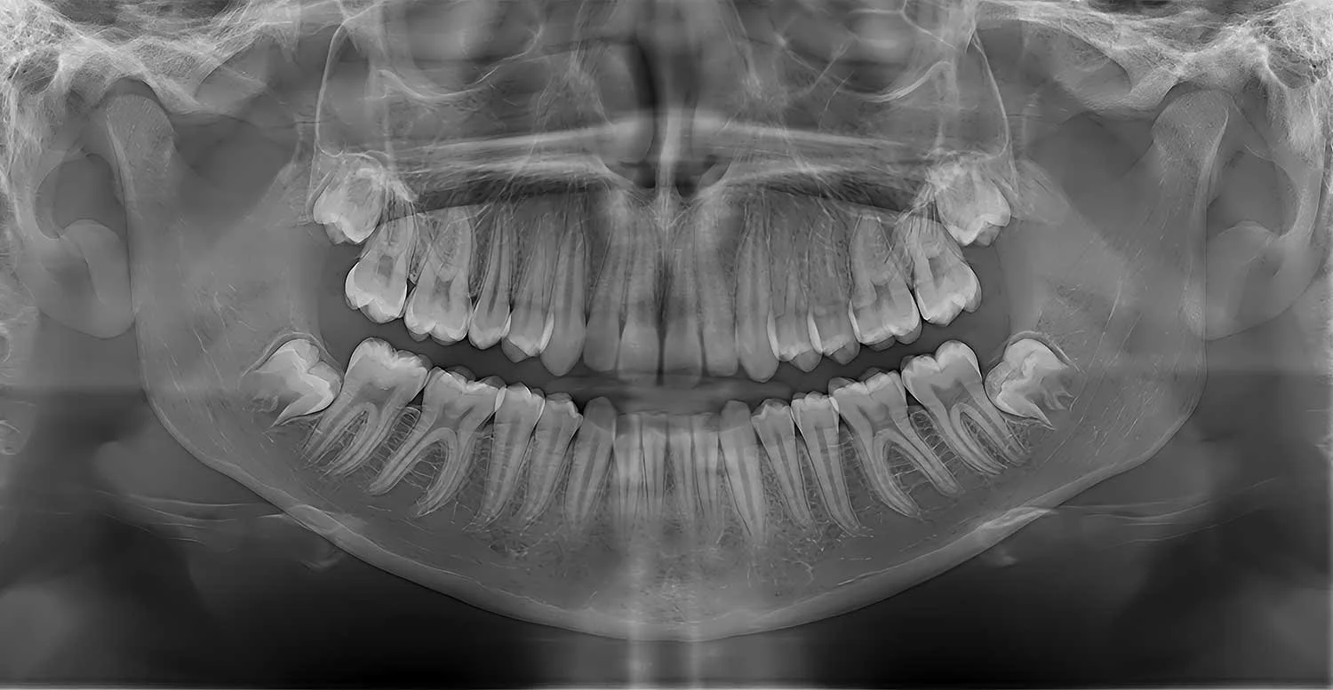
Dental X-rays use ionizing radiation, so it is reasonable to ask why an image is needed and how exposure will be limited. When an X-ray is clinically justified and the equipment and settings are properly selected, the diagnostic benefit is expected to outweigh the small radiation risk. The safest approach is not a fixed schedule for everyone; it is the right image, for the right patient, at the right time.
If you are cautious about radiation, you should be able to have a clear conversation before imaging begins. Your dentist can explain what the image may reveal, whether recent images can be used, and whether a lower-exposure option can answer the same clinical question.
Why Dentists Recommend X Rays
A visual and hands-on dental examination cannot show everything beneath enamel, between teeth, or inside the jaw. X-rays may help identify decay between teeth, bone loss around teeth, infection, impacted teeth, changes near tooth roots, and anatomy needed for treatment planning.
That does not mean every patient needs the same images at every visit. Current ADA and American Academy of Oral and Maxillofacial Radiology recommendations call for a clinical examination first and patient-specific image selection. Age alone, the calendar, or insurance requirements should not be the only reason for an exposure.
How Dental Imaging Exposure Is Reduced
The largest reduction comes from avoiding images that are not needed. Once an image is justified, good technique can lower exposure while still producing an image that is useful for diagnosis.
· Review recent, diagnostically useful images from another dental office when they are available.
· Choose the smallest image type and field of view that can answer the clinical question.
· Use digital receptors and patient-specific exposure settings.
· Restrict the X-ray beam to the area being examined, including rectangular collimation for intraoral images when appropriate.
· Position the patient and receptor carefully to reduce the chance of a retake.
· Use cone-beam computed tomography only when two-dimensional imaging cannot provide the information needed.
Digital imaging can require less exposure than older film systems, but there is no single percentage reduction that applies to every office or every image. Dose depends on the receptor, equipment, beam size, settings, patient size, image type, and technique. For that reason, a specific “80%” or “90% less radiation” promise is less useful than asking how the office selects and optimizes each examination.
Not All Dental Images Have the Same Purpose or Dose
Intraoral 2-D
What it may help evaluate: Cavities between teeth, roots, surrounding bone, or a specific area
Patient-centered question: What finding are you checking for, and are recent images usable?
Panoramic 2-D
What it may help evaluate: A broad view of the jaws, developing or impacted teeth, and selected anatomy
Patient-centered question: Is a broad overview needed, or would a smaller image answer the question?
CBCT 3-D
What it may help evaluate: Three-dimensional anatomy for selected surgical, implant, endodontic, orthodontic, trauma, or pathology questions
Patient-centered question: Why is 3-D information needed, and can the field of view be limited?
What About CBCT Scans
Cone-beam computed tomography creates a three-dimensional view and can answer questions that ordinary dental images cannot. It can be valuable for selected cases, but it generally exposes a patient to more radiation than conventional two-dimensional dental images. It should not be used as a routine screening scan when a lower-exposure examination can provide the information needed.
When CBCT is recommended, ask what clinical question it will answer, whether another image could answer it, and whether the smallest suitable field of view and patient-appropriate settings will be used. Children and adolescents deserve extra care because developing tissues are more sensitive to radiation and they have more years ahead for cumulative effects to matter.
Lead Aprons and Thyroid Collars Have Guidance Changed
Yes. ADA guidance published in 2024 no longer recommends routine lead abdominal aprons or thyroid collars for dental imaging, including during pregnancy. Modern beam restriction and optimized technique protect patients more effectively, while a shield that enters the beam can obscure anatomy and require a retake.
This change can feel counterintuitive, especially if shielding has always represented careful care to you. It does not mean radiation protection matters less. It shifts attention toward preventing unnecessary imaging, limiting the beam, selecting the correct settings, and avoiding retakes. Office procedures must also follow applicable state requirements. If you remain uncomfortable, ask how the recommendation applies to the specific image before it is taken.
Are Dental X Rays Safe During Pregnancy
Dental radiographs are considered safe during pregnancy when they are needed for diagnosis or treatment. Delaying necessary care for pain, infection, or dental disease can create its own risks. Tell the dental team that you are pregnant or may be pregnant so the need for imaging can be reviewed in the context of your health and symptoms.
An image that is not needed now can be deferred. An image needed to diagnose an urgent problem does not have to be postponed simply because of pregnancy. Current ADA guidance does not recommend routine abdominal or thyroid shielding for pregnant dental patients.
A Thoughtful Conversation Before Imaging
Patients should not have to choose between having their concerns dismissed and refusing every X-ray. A respectful decision begins with four questions:
What are you trying to diagnose or rule out?
Do you have recent images that may still be useful?
What is the lowest-exposure examination that can answer this question?
How would the result change today’s treatment decision?
In some situations, declining an image may leave the dentist unable to diagnose a problem or recommend treatment responsibly. If that happens, the dentist should explain what remains unknown, the possible consequences of proceeding without the image, and whether monitoring or another approach is reasonable.
Dental X Ray Safety FAQs
How often should adults get dental X-rays?
There is no single schedule that is right for every adult. The interval depends on current findings, cavity and gum-disease risk, symptoms, treatment history, and whether recent images are available. A patient with active disease may need imaging sooner than a patient with a stable history and low risk.
Are dental X-rays safe during pregnancy?
Yes, dental X-rays can be taken during pregnancy when they are needed for diagnosis or treatment. Tell your dentist that you are pregnant or may be pregnant so the recommendation can be reviewed for your circumstances; routine abdominal and thyroid shielding is no longer recommended by the ADA.
Why do dentists no longer use lead aprons for X-rays?
Current ADA guidance says routine patient shielding is unnecessary with modern, optimized dental imaging. A shield can sometimes block part of the image and cause a retake, while clinical justification, beam restriction, correct settings, and careful positioning reduce exposure more effectively.
Does a dental CBCT scan have more radiation than regular dental X-rays?
In general, CBCT delivers more radiation than conventional two-dimensional dental images, although dose varies widely with the device, settings, and field of view. CBCT should be reserved for a clinical question that cannot be answered adequately with a lower-exposure option.
Talk With Us About Your Imaging Concerns
At Atlanta Dental Wellness, questions about radiation deserve a clear answer. If an image is recommended, ask what it is intended to show and how it may affect your care. If you have recent dental images, arrange to have them sent before your appointment so the dentist can determine whether they remain diagnostically useful.
To discuss your dental health and imaging needs with our Atlanta team, contact Atlanta Dental Wellness or schedule an appointment.